


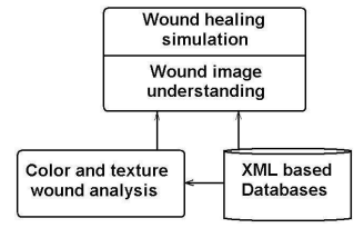
Medical images are valuable in both didactic and research activities, for students inmedicine, pharmacy and health care. Computerized image processing contains methods for non-invasive wound evaluation, allowing an accurate diagnosis in a large category of patients with damaged and wounded skin. Generally, wounds have a non-uniform mixture of yellow slough, red granulation tissue and black necrotic tissue. Relying on a high quality of image acquisition, we can analyze a succession in time of more images for the same wound and assess changes in wound healing, i.e. the recovery or worse evolution. Our aim is to create and implement in Java an automatic method which can be used as a reference standard for colour and texture wound analysis. The purpose is to create e-learning scenarios for wound image understanding and wound healing simulation, by applying this method to large amounts of wound image data stored in XML based knowledge bases (Figure 3.23).
Our main objective is to develop appropriate skills in wound management for a learner that traverses such an e-learning scenario. The e-learning scenarios are practice driven and relevant to professional practice, being used by students in medicine, pharmacy and healthcare, at graduate, postgraduate and residency levels.
For a given wound, we must find out some quantitative and qualitative attributes for assessing the healing state. As quantitative attributes we measure its surface area and its volume (evaluating depth). The original image is processed with the purpose to emphasize the distinction between wound and non-wound area. We use some general methods to enhance the image, because we must exaggerate the distinction between wound and non-wound. As an example, for individuals with fair skin, we lighten the images and then view them using shades of green with the red and blue minimized. This way more clearly exhibit the borders of the wound than in the original image. Removal of the red and blue leaves the wound black and the rest of the image green. For images of individuals with dark skin, both the red and green are accentuated while the blue is minimized. This procedure also leaves the non-wound area green, but colours the wound red. In either case, the wound can easily be distinguished from the non-wound without difficulty.
Our work is based on a continuous collaboration with physicians and wound care experts, because it is necessary to make a rigorous classification for various categories of wounds. We collected large amounts of wound image data and we calculate statistical parameters as mean, median, standard deviation, confidence interval, skewness and kurtosis for them. These historical data are included in XML based databases, to be used as inputs to classification algorithms. The general purpose is to make distinction between infected and non-infected, inflamed and non-inflamed wounds. Based on colour analysis, we build a statistically significant differentiation of mild, moderate and severe wounds. Our system analyses the differences in calibrated hue between injured and non-injured skin, obtaining a repeatable differentiation of wound severity for various time intervals. As an example, burn wounds are characterized according to their depth as:
- Superficial – with bright red colour and the presence of blisters (usually with brown colour);
- Deep – with red-whitish colour and with dark dots;
- Deep – with red-whitish colour and with dark dots;
Assuming normality, the first two moments (mean and standard deviation) characterize very well the colour distribution. However, sometimes the first two moments alone are inadequate to discriminate between wound and non-wound skin. Therefore further details of the colour distribution are required. Skewness and kurtosis of the colour data proved to be more useful for this purpose.
We implement e-tools that will enable to assess the current state of the wound and to gain insight into the wound evolution, by comparing the series of wound data collected over time. Based on this knowledge we can design an e-tool for simulating the process of wound healing. The colour image processing is the most acceptable automatic method of objectively and reproducibly analyzing skin wounds and lesions. Also, this method has the main advantage of being completely non-invasive.
- 1879 reads
