


When the German physicists Gustav Fritsch and Eduard Hitzig (1870/2009) 1 applied mild electric stimulation to different parts of a dog’s cortex, they discovered that they could make different parts of the dog’s body move. Furthermore, they discovered an important and unexpected principle of brain activity. They found that stimulating the right side of the brain produced movement in the left side of the dog’s body, and vice versa. This finding follows from a general principle about how the brain is structured, called contralateral control. The brain is wired such that in most cases the left hemisphere receives sensations from and controls the right side of the body, and vice versa.
Fritsch and Hitzig also found that the movement that followed the brain stimulation only occurred when they stimulated a specific arch-shaped region that runs across the top of the brain from ear to ear, just at the front of the parietal lobe (see Figure 3.9). Fritsch and Hitzig had discovered the motor cortex, the part of the cortex that controls and executes movements of the body by sending signals to the cerebellum and the spinal cord. More recent research has mapped the motor cortex even more fully, by providing mild electronic stimulation to different areas of the motor cortex in fully conscious patients while observing their bodily responses (because the brain has no sensory receptors, these patients feel no pain). As you can see in Figure 3.9, this research has revealed that the motor cortex is specialized for providing control over the body, in the sense that the parts of the body that require more precise and finer movements, such as the face and the hands, also are allotted the greatest amount of cortical space.
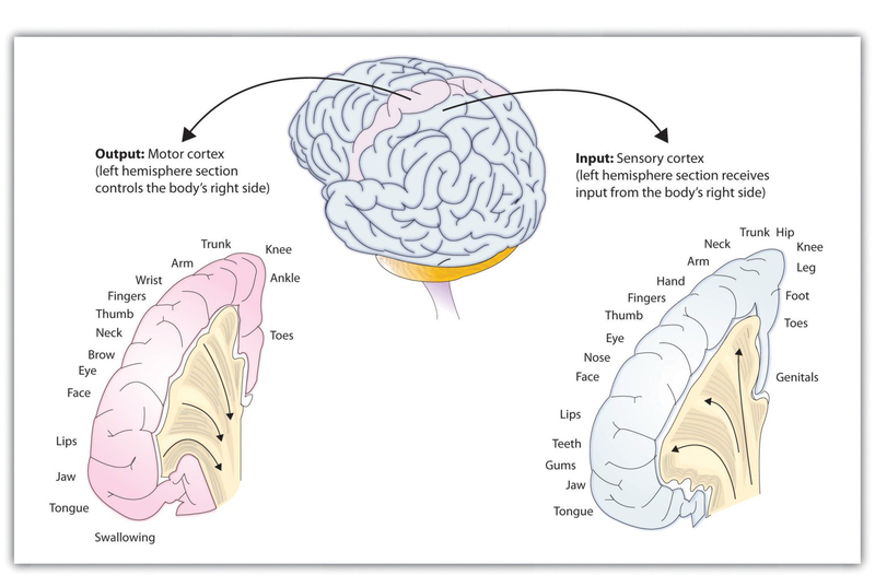
Just as the motor cortex sends out messages to the specific parts of the body, the somatosensory cortex, an area just behind and parallel to the motor cortex at the back of the frontal lobe, receives information from the skin’s sensory receptors and the movements of different body parts. Again, the more sensitive the body region, the more area is dedicated to it in the sensory cortex. Our sensitive lips, for example, occupy a large area in the sensory cortex, as do our fingers and genitals.
Other areas of the cortex process other types of sensory information. The visual cortex is the area located in the occipital lobe (at the very back of the brain) that processes visual information. If you were stimulated in the visual cortex, you would see flashes of light or color, and perhaps you remember having had the experience of “seeing stars” when you were hit in, or fell on, the back of your head. The temporal lobe, located on the lower side of each hemisphere, contains the auditory cortex, which is responsible for hearing and language. The temporal lobe also processes some visual information, providing us with the ability to name the objects around us (Martin, 2007). 2
As you can see in Figure 3.9, the motor and sensory areas of the cortex account for a relatively small part of the total cortex. The remainder of the cortex is made up of association areas in which sensory and motor information is combined and associated with our stored knowledge. These association areas are the places in the brain that are responsible for most of the things that make human beings seem human. The association areas are involved in higher mental functions, such as learning, thinking, planning, judging, moral reflecting, figuring, and spatial reasoning.
- 5477 reads
