


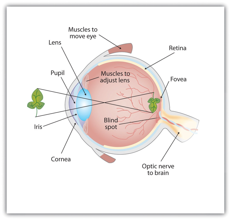
As you can see in Figure 4.4, light enters the eye through the cornea, a clear covering that protects the eye and begins to focus the incoming light. The light then passes through the pupil, a small opening in the center of the eye. The pupil is surrounded by the iris, the colored part of the eye that controls the size of the pupil by constricting or dilating in response to light intensity. When we enter a dark movie theater on a sunny day, for instance, muscles in the iris open the pupil and allow more light to enter. Complete adaptation to the dark may take up to 20 minutes.
Behind the pupil is the lens, a structure that focuses the incoming light on the retina, the layer of tissue at the back of the eye that contains photoreceptor cells. As our eyes move from near objects to distant objects, a process known as visual accommodation occurs. Visual accommodation is the process of changing the curvature of the lens to keep the light entering the eye focused on the retina. Rays from the top of the image strike the bottom of the retina and vice versa, and rays from the left side of the image strike the right part of the retina and vice versa, causing the image on the retina to be upside down and backward. Furthermore, the image projected on the retina is flat, and yet our final perception of the image will be three dimensional.
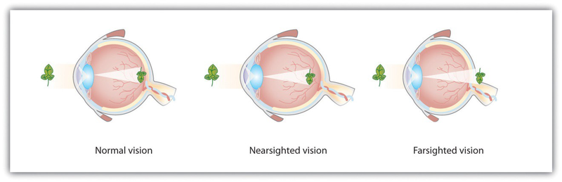
Accommodation is not always perfect, and in some cases the light that is hitting the retina is a bit out of focus. As you can see in Figure 4.5, if the focus is in front of the retina, we say that the person is nearsighted, and when the focus is behind the retina we say that the person is farsighted. Eyeglasses and contact lenses correct this problem by adding another lens in front of the eye, and laser eye surgery corrects the problem by reshaping the eye’s own lens.
The retina contains layers of neurons specialized to respond to light (see Figure 4.6). As light falls on the retina, it first activates receptor cells known as rods and cones. The activation of these cells then spreads to the bipolar cells and then to the ganglion cells, which gather together and converge, like the strands of a rope, forming the optic nerve. The optic nerve is a collection of millions of ganglion neurons that sends vast amounts of visual information, via the thalamus, to the brain. Because the retina and the optic nerve are active processors and analyzers of visual information, it is not inappropriate to think of these structures as an extension of the brain itself.
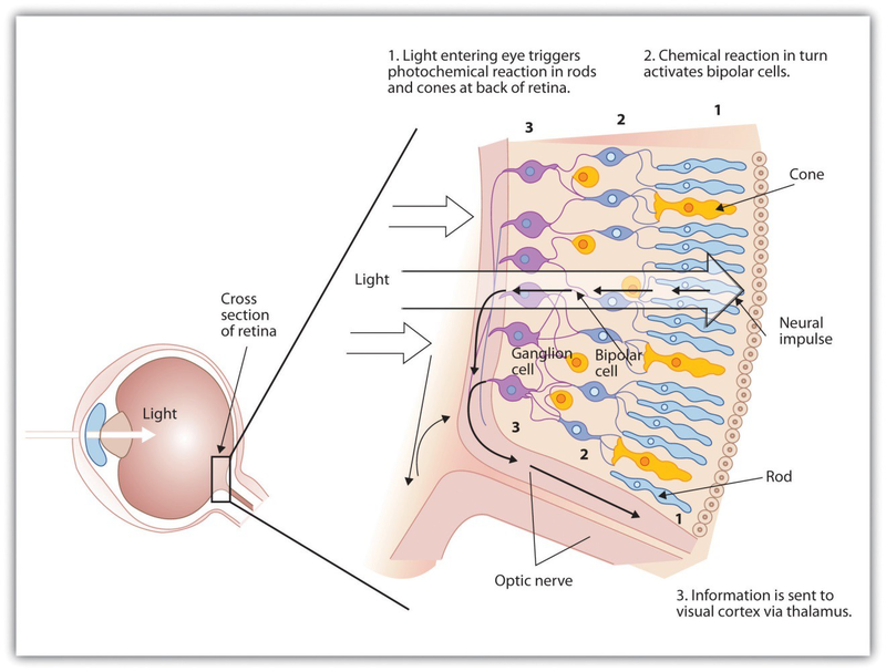
Rods are visual neurons that specialize in detecting black, white, and gray colors. There are about 120 million rods in each eye. The rods do not provide a lot of detail about the images we see, but because they are highly sensitive to shorter-waved (darker) and weak light, they help us see in dim light, for instance, at night. Because the rods are located primarily around the edges of the retina, they are particularly active in peripheral vision (when you need to see something at night, try looking away from what you want to see). Cones are visual neurons that are specialized in detecting fine detail and colors. The 5 million or so cones in each eye enable us to see in color, but they operate best in bright light. The cones are located primarily in and around the fovea, which is the central point of the retina.
To demonstrate the difference between rods and cones in attention to detail, choose a word in this text and focus on it. Do you notice that the words a few inches to the side seem more blurred? This is because the word you are focusing on strikes the detail-oriented cones, while the words surrounding it strike the less-detail-oriented rods, which are located on the periphery.
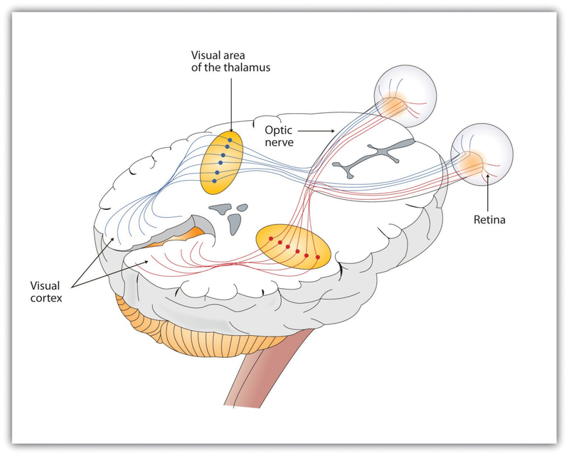
As you can see in Figure 4.7, the sensory information received by the retina is relayed through the thalamus to corresponding areas in the visual cortex, which is located in the occipital lobe at the back of the brain. Although the principle of contralateral control might lead you to expect that the left eye would send information to the right brain hemisphere and vice versa, nature is smarter than that. In fact, the left and right eyes each send information to both the left and the right hemisphere, and the visual cortex processes each of the cues separately and in parallel. This is an adaptational advantage to an organism that loses sight in one eye, because even if only one eye is functional, both hemispheres will still receive input from it.
The visual cortex is made up of specialized neurons that turn the sensations they receive from the optic nerve into meaningful images. Because there are no photoreceptor cells at the place where the optic nerve leaves the retina, a hole or blind spot in our vision is created (see Figure 4.8). When both of our eyes are open, we don’t experience a problem because our eyes are constantly moving, and one eye makes up for what the other eye misses. But the visual system is also designed to deal with this problem if only one eye is open—the visual cortex simply fills in the small hole in our vision with similar patterns from the surrounding areas, and we never notice the difference. The ability of the visual system to cope with the blind spot is another example of how sensation and perception work together to create meaningful experience.
Perception is created in part through the simultaneous action of thousands of feature detector neurons—specialized neurons, located in the visual cortex, that respond to the strength, angles, shapes, edges, and movements of a visual stimulus (Kelsey, 1997; Livingstone & Hubel, 1988). 1 The feature detectors work in parallel, each performing a specialized function. When faced with a red square, for instance, the parallel line feature detectors, the horizontal line feature detectors, and the red color feature detectors all become activated. This activation is then passed on to other parts of the visual cortex where other neurons compare the information supplied by the feature detectors with images stored in memory. Suddenly, in a flash of recognition, the many neurons fire together, creating the single image of the red square that we experience (Rodriguez et al., 1999). 2
Some feature detectors are tuned to selectively respond to particularly important objects, for instance, faces, smiles, and other parts of the body (Downing, Jiang, Shuman, & Kanwisher, 2001; Haxby et al., 2001). 3 When researchers disrupted face recognition areas of the cortex using the magnetic pulses of transcranial magnetic stimulation (TMS), people were temporarily unable to recognize faces, and yet they were still able to recognize houses (McKone, Kanwisher,& Duchaine, 2007; Pitcher, Walsh, Yovel, & Duchaine, 2007). 4
- 5957 reads
