


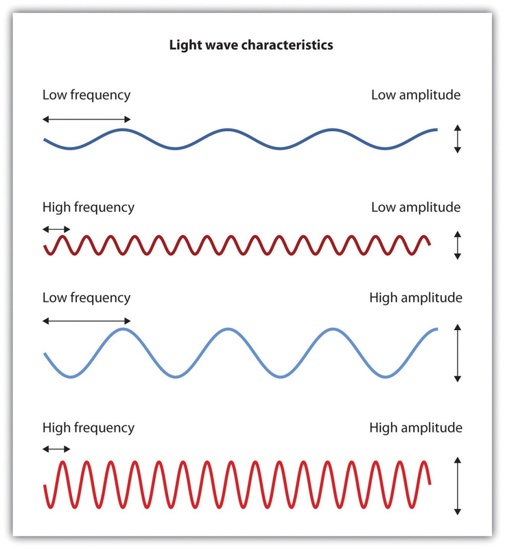
It has been estimated that the human visual system can detect and discriminate among 7 million color variations (Geldard, 1972), 1 but these variations are all created by the combinations of the three primary colors: red, green, and blue. The shade of a color, known as hue, is conveyed by the wavelength of the light that enters the eye (we see shorter wavelengths as more blue and longer wavelengths as more red), and we detect brightness from the intensity or height of the wave (bigger or more intense waves are perceived as brighter).
In his important research on color vision, Hermann von Helmholtz (1821–1894) theorized that color is perceived because the cones in the retina come in three types. One type of cone reacts primarily to blue light (short wavelengths), another reacts primarily to green light (medium wavelengths), and a third reacts primarily to red light (long wavelengths). The visual cortex then detects and compares the strength of the signals from each of the three types of cones, creating the experience of color. According to this Young-Helmholtz trichromatic color theory, what color we see depends on the mix of the signals from the three types of cones. If the brain is receiving primarily red and blue signals, for instance, it will perceive purple; if it is receiving primarily red and green signals it will perceive yellow; and if it is receiving messages from all three types of cones it will perceive white.
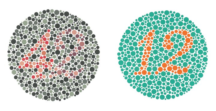
The different functions of the three types of cones are apparent in people who experience color blindness—the inability to detect either green and/or red colors. About 1 in 50 people, mostly men, lack functioning in the red- or green-sensitive cones, leaving them only able to experience either one or two colors (Figure 4.11).
The trichromatic color theory cannot explain all of human vision, however. For one, although the color purple does appear to us as a mixing of red and blue, yellow does not appear to be a mix of red and green. And people with color blindness, who cannot see either green or red, nevertheless can still see yellow. An alternative approach to the Young-Helmholtz theory, known as the opponent-process color theory, proposes that we analyze sensory information not in terms of three colors but rather in three sets of “opponent colors”: red-green, yellow-blue, and white- black. Evidence for the opponent-process theory comes from the fact that some neurons in the retina and in the visual cortex are excited by one color (e.g., red) but inhibited by another color (e.g., green).
One example of opponent processing occurs in the experience of an afterimage. If you stare at the flag on the left side of Figure 4.12 for about 30 seconds (the longer you look, the better the effect), and then move your eyes to the blank area to the right of it, you will see the afterimage. When we stare at the green stripes, our green receptors habituate and begin to process less strongly, whereas the red receptors remain at full strength. When we switch our gaze, we see primarily the red part of the opponent process. Similar processes create blue after yellow and white after black.

The tricolor and the opponent-process mechanisms work together to produce color vision. When light rays enter the eye, the red, blue, and green cones on the retina respond in different degrees, and send different strength signals of red, blue, and green through the optic nerve. The color signals are then processed both by the ganglion cells and by the neurons in the visual cortex (Gegenfurtner & Kiper, 2003). 2
- 3365 reads
