


Still another approach to measuring thoughts and feelings is to measure brain activity, and recent advances in brain science have created a wide variety of new techniques for doing so. One approach, known as electroencephalography (EEG), is a technique that records the electrical activity produced by the brain’s neurons through the use of electrodes that are placed around the research participant’s head. An electroencephalogram (EEG) can show if a person is asleep, awake, or anesthetized because the brain wave patterns are known to differ during each state. An EEG can also track the waves that are produced when a person is reading, writing, and speaking with others. A particular advantage of the technique is that the participant can move around while the recordings are being taken, which is useful when measuring brain activity in children who often have difficulty keeping still. Furthermore, by following electrical impulses across the surface of the brain, researchers can observe changes over very fast time periods.
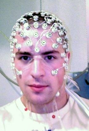
Although EEGs can provide information about the general patterns of electrical activity within the brain, and although they allow the researcher to see these changes quickly as they occur in real time, the electrodes must be placed on the surface of the skull, and each electrode measures brain waves from large areas of the brain. As a result, EEGs do not provide a very clear picture of the structure of the brain.
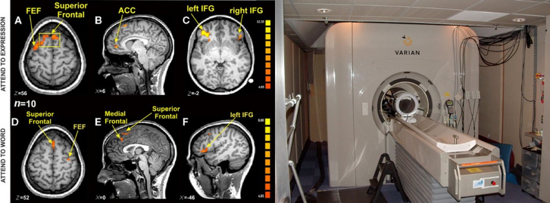
But techniques exist to provide more specific brain images. Functional magnetic resonance imaging (fMRI) is a neuroimaging technique that uses a magnetic field to create images of brain structure and function. In research studies that use the fMRI, the research participant lies on a bed within a large cylindrical structure containing a very strong magnet. Nerve cells in the brain that are active use more oxygen, and the need for oxygen increases blood flow to the area. The fMRI detects the amount of blood flow in each brain region and thus is an indicator of which parts of the brain are active.
Very clear and detailed pictures of brain structures (see Figure 1.9) can be produced via fMRI. Often, the images take the form of cross-sectional “slices” that are obtained as the magnetic field is passed across the brain. The images of these slices are taken repeatedly and are superimposed on images of the brain structure itself to show how activity changes in different brain structures over time. Normally, the research participant is asked to engage in tasks while in the scanner, for instance, to make judgments about pictures of people, to solve problems, or to make decisions about appropriate behaviors. The fMRI images show which parts of the brain are associated with which types of tasks. Another advantage of the fMRI is that is it noninvasive. The research participant simply enters the machine and the scans begin.
Although the scanners themselves are expensive, the advantages of fMRIs are substantial, and scanners are now available in many university and hospital settings. The fMRI is now the most commonly used method of learning about brain structure, and it has been employed by social psychologists to study social cognition, attitudes, morality, emotions, responses to being rejected by others, and racial prejudice, to name just a few topics (Eisenberger, Lieberman, & Williams, 2003; Greene, Sommerville, Nystrom, Darley, & Cohen, 2001; Lieberman, Hariri, Jarcho, Eisenberger, & Bookheimer, 2005; Ochsner, Bunge, Gross, & Gabrieli, 2002; Richeson et al., 2003).
- 3348 reads
