


Take a breath in and hold it. Wait several seconds and then let it out. Humans, when they are not exerting themselves, breathe approximately 15 times per minute on average. This equates to about 900 breaths an hour or 21,600 breaths per day. With every inhalation, air fills the lungs, and with every exhalation, it rushes back out. That air is doing more than just inflating and deflating the lungs in the chest cavity. The air contains oxygen that crosses the lung tissue, enters the bloodstream, and travels to organs and tissues. There, oxygen is exchanged for carbon dioxide, which is a cellular waste material. Carbon dioxide exits the cells, enters the bloodstream, travels back to the lungs, and is expired out of the body during exhalation.
Breathing is both a voluntary and an involuntary event. How often a breath is taken and how much air is inhaled or exhaled is regulated by the respiratory center in the brain in response to signals it receives about the carbon dioxide content of the blood. However, it is possible to override this automatic regulation for activities such as speaking, singing and swimming under water.
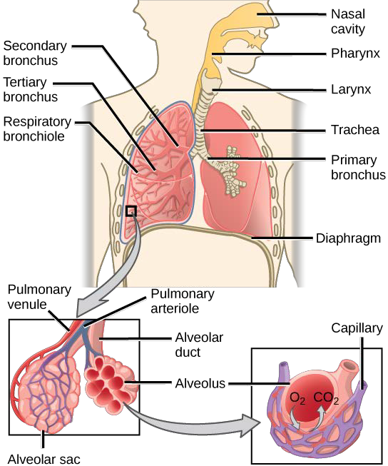
During inhalation the diaphragm descends creating a negative pressure around the lungs and they begin to inflate, drawing in air from outside the body. The air enters the body through the nasalcavity located just inside the nose (Figure 16.9). As the air passes through the nasal cavity, the air is warmed to body temperature and humidified by moisture from mucous membranes. These processes help equilibrate the air to the body conditions, reducing any damage that cold, dry air can cause. Particulate matter that is floating in the air is removed in the nasal passages by hairs, mucus, and cilia. Air is also chemically sampled by the sense of smell.
From the nasal cavity, air passes through the pharynx (throat) and the larynx (voice box) as it makes its way to the trachea (Figure 16.9). The main function of the trachea is to funnel the inhaled air to the lungs and the exhaled air back out of the body. The human trachea is a cylinder, about 25 to 30 cm (9.8–11.8 in) long, which sits in front of the esophagus and extends from the pharynx into the chest cavity to the lungs. It is made of incomplete rings of cartilage and smooth muscle. The cartilage provides strength and support to the trachea to keep the passage open. The trachea is lined with cells that have cilia and secrete mucus. The mucus catches particles that have been inhaled, and the cilia move the particles toward the pharynx.
The end of the trachea divides into two bronchi that enter the right and left lung. Air enters the lungs through the primary bronchi. The primary bronchus divides, creating smaller and smaller diameter bronch iuntil the passages are under 1 mm (.03 in) in diameter when they are called bronchioles as they split and spread through the lung. Like the trachea, the bronchus and bronchioles are made of cartilage and smooth muscle. Bronchi are innervated by nerves of both the parasympathetic and sympathetic nervous systems that control muscle contraction (parasympathetic) or relaxation (sympathetic) in the bronchi and bronchioles, depending on the nervous system’s cues. The final bronchioles are the respiratory bronchioles. Alveolar ducts are attached to the end of each respiratory bronchiole. At the end of each duct are alveolar sacs, each containing 20 to 30 alveoli. Gas exchange occurs only in the alveoli. The alveoli are thin-walled and look like tiny bubbles within the sacs. The alveoli are in direct contact with capillaries of the circulatory system. Such intimate contact ensures that oxygen will diffuse from the alveoli into the blood. In addition, carbon dioxide will diffuse from the blood into the alveoli to be exhaled. The anatomical arrangement of capillaries and alveoli emphasizes the structural and functional relationship of the respiratory and circulatory systems. Estimates for the surface area of alveoli in the lungs vary around 100 m2. This large area is about the area of half a tennis court. This large surface area, combined with the thin-walled nature of the alveolar cells, allows gases to easily diffuse across the cells.
Art CONNECTION
Which of the following statements about the human respiratory system is false?
- When we breathe in, air travels from the pharynx to the trachea.
- The bronchioles branch into bronchi.
- Alveolar ducts connect to alveolar sacs.
- Gas exchange between the lungs and blood takes place in the alveolus.

Watch this video (http://openstaxcollege.org/l/lungs_pulmonar2) for a review of the respiratory system.
- 4342 reads
