


The endocrine glands secrete hormones into the surrounding interstitial fluid; those hormones then diffuse into blood and are carried to various organs and tissues within the body. The endocrine glands include the pituitary, thyroid, parathyroid, adrenal glands, gonads, pineal, and pancreas.
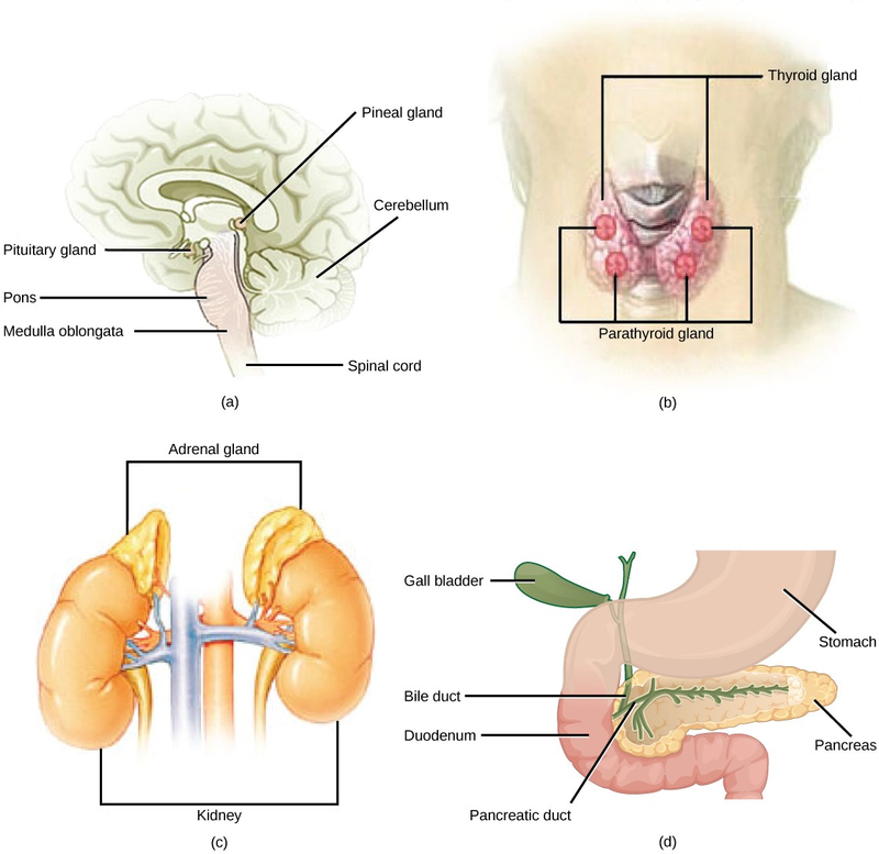
The pituitary gland, sometimes called the hypophysis, is located at the base of the brain (Figure 16.13). It is attached to the hypothalamus. The posterior lobe stores and releases oxytocin and antidiuretic hormone produced by the hypothalamus. The anterior lobe responds to hormones produced by the hypothalamus by producing its own hormones, most of which regulate other hormone-producing glands.
The anterior pituitary produces six hormones: growth hormone, prolactin, thyroid-stimulating hormone, adrenocorticotropic hormone, follicle-stimulating hormone, and luteinizing hormone. Growth hormone stimulates cellular activities like protein synthesis that promote growth. Prolactin stimulates the production of milk by the mammary glands. The other hormones produced by the anterior pituitary regulate the production of hormones by other endocrine tissues (Table 16.1). The posterior pituitary is significantly different in structure from the anterior pituitary. It is a part of the brain, extending down from the hypothalamus, and contains mostly nerve fibers that extend from the hypothalamus to the posterior pituitary.
The thyroid gland is located in the neck, just below the larynx and in front of the trachea (Figure 16.13). It is a butterfly-shaped gland with two lobes that are connected. The thyroid follicle cells synthesize the hormone thyroxine, which is also known as T4 because it contains four atoms of iodine, and triiodothyronine, also known as T3 because it contains three atoms of iodine. T3 and T4 are released by the thyroid in response to thyroid-stimulating hormone produced by the anterior pituitary, and both T3 and T4 have the effect of stimulating metabolic activity in the body and increasing energy use. A third hormone, calcitonin, is also produced by the thyroid. Calcitonin is released in response to rising calcium ion concentrations in the blood and has the effect of reducing those levels.
Most people have four parathyroid glands; however, the number can vary from two to six. These glands are located on the posterior surface of the thyroid gland (Figure 16.13).
The parathyroid glands produce parathyroid hormone. Parathyroid hormone increases blood calcium concentrations when calcium ion levels fall below normal.
The adrenal glands are located on top of each kidney (Figure 16.13). The adrenal glands consist of an outer adrenal cortex and an inner adrenal medulla. These regions secrete different hormones.
The adrenal cortex produces mineralocorticoids, glucocorticoids, and androgens. The main mineralocorticoid is aldosterone, which regulates the concentration of ions in urine, sweat, and saliva. Aldosterone release from the adrenal cortex is stimulated by a decrease in blood concentrations of sodium ions, blood volume, or blood pressure, or by an increase in blood potassium levels. The glucocorticoids maintain proper blood-glucose levels between meals. They also control a response to stress by increasing glucose synthesis from fats and proteins and interact with epinephrine to cause vasoconstriction. Androgens are sex hormones that are produced in small amounts by the adrenal cortex. They do not normally affect sexual characteristics and may supplement sex hormones released from the gonads. The adrenal medulla contains two types of secretory cells: one that produces epinephrine (adrenaline) and another that produces norepinephrine (noradrenaline). Epinephrine and norepinephrine cause immediate, short-term changes in response to stressors, inducing the so-called fight-or-flight response. The responses include increased heart rate, breathing rate, cardiac muscle contractions, and blood-glucose levels. They also accelerate the breakdown of glucose in skeletal muscles and stored fats in adipose tissue, and redirect blood flow toward skeletal muscles and away from skin and viscera. The release of epinephrine and norepinephrine is stimulated by neural impulses from the sympathetic nervous system that originate from the hypothalamus.
The pancreas is an elongate organ located between the stomach and the proximal portion of the small intestine (Figure 16.13). It contains both exocrine cells that excrete digestive enzymes and endocrine cells that release hormones.
The endocrine cells of the pancreas form clusters called pancreatic islets or the islets of Langerhans. Among the cell types in each pancreatic islet are the alpha cells, which produce the hormone glucagon, and the beta cells, which produce the hormone insulin. These hormones regulate blood-glucose levels. Alpha cells release glucagon as blood-glucose levels decline. When blood-glucose levels rise, beta cells release insulin. Glucagon causes the release of glucose to the blood from the liver, and insulin facilitates the uptake of glucose by the body’s cells.
The gonads—the male testes and female ovaries—produce steroid hormones. The testes produce androgens, testosterone being the most prominent, which allow for the development of secondary sex characteristics and the production of sperm cells. The ovaries produce estrogen and progesterone, which cause secondary sex characteristics, regulate production of eggs, control pregnancy, and prepare the body for childbirth.
There are several organs whose primary functions are non-endocrine but that also possess endocrine functions. These include the heart, kidneys, intestines, thymus, and adipose tissue. The heart has endocrine cells in the walls of the atria that release a hormone in response to increased blood volume. It causes a reduction in blood volume and blood pressure, and reduces the concentration of Na+ in the blood.
The gastrointestinal tract produces several hormones that aid in digestion. The endocrine cells are located in the mucosa of the GI tract throughout the stomach and small intestine. They trigger the release of gastric juices, which help to break down and digest food in the GI tract.
The kidneys also possess endocrine function. Two of these hormones regulate ion concentrations and blood volume or pressure. Erythropoietin (EPO) is released by kidneys in response to low oxygen levels. EPO triggers the formation of red blood cells in the bone marrow. EPO has been used by athletes to improve performance. But EPO doping has its risks, since it thickens the blood and increases strain on the heart; it also increases the risk of blood clots and therefore heart attacks and stroke.
The thymus is found behind the sternum. The thymus produces hormones referred to as thymosins, which contribute to the development of the immune response in infants. Adipose tissue, or fat tissue, produces the hormone leptin in response to food intake. Leptin produces a feeling of satiety after eating, reducing the urge for further eating.
|
Endocrine Associated Gland Hormones Effect |
||
|
Pituitary (anterior) |
growth hormone | promotes growth of body tissues |
| prolactin | promotes milk production | |
|
thyroid-stimulating hormone |
stimulates thyroid hormone release |
|
|
adrenocorticotropic hormone |
stimulates hormone release by adrenal cortex |
|
|
follicle-stimulating hormone |
stimulates gamete production |
|
|
luteinizing hormone |
stimulates androgen production by gonads in males; stimulates ovulation and production of estrogen and progesterone in females |
|
|
Pituitary (posterior) |
antidiuretic hormone |
stimulates water reabsorption by kidneys |
|
oxytocin stimulates uterine contractions during childbirth |
||
|
Thyroid |
thyroxine, triiodothyronine |
stimulate metabolism |
|
calcitonin reduces blood Ca2+ levels |
||
|
Parathyroid |
parathyroid hormone |
increases blood Ca2+ levels |
|
Adrenal (cortex) |
aldosterone increases blood Na+ levels |
|
|
cortisol, corticosterone, cortisone |
increase blood-glucose levels |
|
|
Adrenal (medulla) |
epinephrine, norepinephrine |
stimulate fight-or-flight response |
|
Pancreas |
insulin reduces blood-glucose levels |
|
|
glucagon |
increases blood-glucose levels |
|
- 2660 reads
