


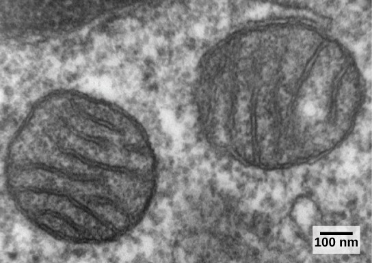
Eukaryotic cells may contain anywhere from one to several thousand mitochondria, depending on the cell’s level of energy consumption. Each mitochondrion measures 1 to 10 micrometers in length and exists in the cell as a moving, fusing, and dividing oblong spheroid (Figure 13.11). However, mitochondria cannot survive outside the cell. As the atmosphere was oxygenated by photosynthesis, and as successful aerobic prokaryotes evolved, evidence suggests that an ancestral cell engulfed and kept alive a free-living, aerobic prokaryote. This gave the host cell the ability to use oxygen to release energy stored in nutrients. Several lines of evidence support that mitochondria are derived from this endosymbiotic event. Mitochondria are shaped like a specific group of bacteria and are surrounded by two membranes, which would result when one membrane-bound organism was engulfed by another membrane-bound organism. The mitochondrial inner membrane involves substantial infoldings or cristae that resemble the textured outer surface of certain bacteria.
Mitochondria divide on their own by a process that resembles binary fission in prokaryotes. Mitochondria have their own circular DNA chromosome that carries genes similar to those expressed by bacteria. Mitochondria also have special ribosomes and transfer RNAs that resemble these components in prokaryotes. These features all support that mitochondria were once free-living prokaryotes.
- 2819 reads
