


Fertilization is the process in which gametes (an egg and sperm) fuse to form a zygote (Figure 18.8). To ensure that the offspring has only one complete diploid set of chromosomes, only one sperm must fuse with one egg. In mammals, a layer called the zona pellucida protects the egg. At the tip of the head of a sperm cell is a structure like a lysosome called the acrosome, which contains enzymes. When a sperm binds to the zona pellucida, a series of events, called the acrosomal reactions, take place. These reactions, involving enzymes from the acrosome, allow the sperm plasma membrane to fuse with the egg plasma membrane and permit the sperm nucleus to transfer into the ovum. The nuclear membranes of the egg and sperm break down and the two haploid nuclei fuse to form a diploid nucleus or genome.
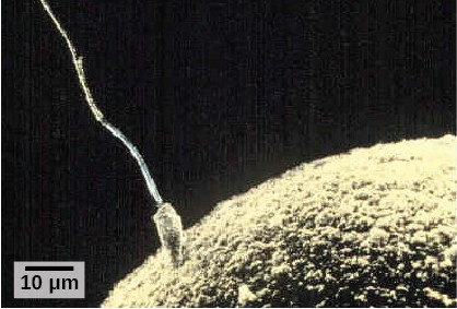
To ensure that no more than one sperm fertilizes the egg, once the acrosomal reactions take place at one location of the egg membrane, the egg releases proteins in other locations to prevent other sperm from fusing with the egg.
The development of multi-cellular organisms begins from this single-celled zygote, which undergoes rapid cell division, called cleavage (Figure 18.9), to form a hollow ball of cells called a blastula Figure 18.9).
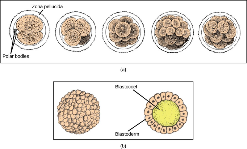
In mammals, the blastula forms the blastocystin the next stage of development. Here the cells in the blastula arrange themselves in two layers: the innercellmass, and an outer layer called the trophoblast. The inner cell mass will go on to form the embryo. The trophoblast secretes enzymes that allow implantation of the blastocyst into the endometrium of the uterus. The trophoblast will contribute to the placenta and nourish the embryo.

Visit the Virtual Human Embryo project (http://openstaxcollege.org/l/human_embryo2) at the Endowment for Human Development site to click through an interactive of the stages of embryo development, including micrographs and rotating 3-D images.
The cells in the blastula then rearrange themselves spatially to form three layers of cells. This process is called gastrulation. During gastrulation, the blastula folds in on itself and cells migrate to form the three layers of cells (Figure 18.10) in a structure, the gastrula, with a hollow space that will become the digestive tract. Each of the layers of cells is called a germ layer and will differentiate into different organ systems.
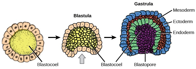
The three germ layers are the endoderm, the ectoderm, and the mesoderm. Cells in each germ layer differentiate into tissues and embryonic organs. The ectoderm gives rise to the nervous system and the epidermis, among other tissues. The mesoderm gives rise to the muscle cells and connective tissue in the body. The endoderm gives rise to the gut and many internal organs.
- 2903 reads
